Abstract

Introduction: Patients with chronic anemia syndromes, such as thalassemia major and sickle cell disease, have premature white matter volume loss in characteristic watershed areas, suggesting ischemic damage. We have previously demonstrated that resting brain blood flow is increased in these patients, suggesting adequate oxygen delivery at rest. However, we postulate that increased blood flow velocity in the microvascular beds might not provide adequate time for oxygen exchange, leading to decreased arterio-venous oxygen gradients. To test this hypothesis, we evaluated peripheral (brachial) and cerebral arteriovenous oxygen gradients in 24 patients with chronic anemia syndromes and 45 control subjects.
Methods: All patients were part of a larger study evaluating cerebral blood flow and oxygen delivery in chronic anemia syndromes. For the present comparison, we excluded patients with sickle cell anemia because sickle cells have abnormal magnetic properties. Anemic subjects included 15 patients with thalassemia major, three patients with E-beta thalassemia, three patients with hemoglobin H-constant spring, two patients with hereditary spherocytosis, and one patient with dyserythropoetic anemia. 45 age matched healthy subjects were recruited from friends and family of the patient cohort. Peripheral oxygen saturation was measured by fingertip pulse oximetry. Brachial venous oxygen saturation was assessed by cooximetry (blood gas was drawn after tourniquet release). Sagittal sinus oxygen saturation was estimated using T2 Relaxation Under Spin Tagging (TRUST), a validated MRI technique for noninvasive oximetry in the brain.
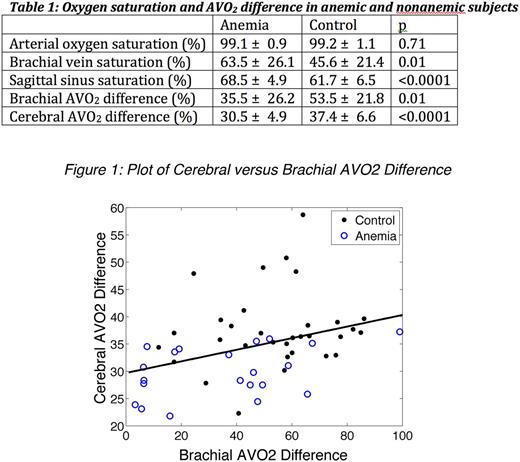
Results: Table 1 summarizes the oxygen saturation and arteriovenous oxygen gradients (AVO2 differences) between the two populations. No difference was observed in arterial oxygen saturation, however both brachial and sagittal sinus saturations were significantly higher in anemic patients. AVO2 gradients were markedly lower in anemic subjects, indicating lower oxygen extraction in the periphery as well as the brain. AVO2 gradients were correlated with hemoglobin (r2 = 0.17, p=0.002) in both the brain and periphery. AVO2 gradient in the brain was correlated with AVO2 gradient in the forearm (Figure 1).
Discussion: We demonstrate that arteriovenous oxygen gradients are decreased in anemic subjects, relative to controls, in both peripheral and cerebral vascular beds. Cardiac output and cerebral blood flow are increased in anemic subjects to preserve oxygen delivery. However, increased blood flow potentially shortens microvascular transit time, decreasing the oxygen exchange time. It is not yet clear whether the lower AVO2 gradients are inherently pathologic, but clearly represent an abnormal physiology with potential impact on the white matter disease observed in these patients.
No relevant conflicts of interest to declare.
Author notes
Asterisk with author names denotes non-ASH members.

This icon denotes a clinically relevant abstract


This feature is available to Subscribers Only
Sign In or Create an Account Close Modal